Health of the French bulldog.
Brachycephalic dogs can suffer with numerous health conditions…..
BOAS - Brachycephalic Obstructive Airway Syndrome
Buying a pup? Don't take the words of "health checked parents" to qualify that they have had the proper testing done along with BOAS scoring **
Why is my dog not ‘cured’ by BOAS surgery?
Surgery cannot cure BOAS but instead aims to alleviate the symptoms and reduce the likelihood of further deterioration.
BOAS is a complicated syndrome resulting from multiple abnormalities within the upper and lower airways. Some of these abnormalities can be improved with surgery (e.g. narrow nostrils, overly long soft palate) but others are either impossible or very difficult to treat (e.g. hypoplastic trachea, oversized tongue and thick soft palate). Some dogs also have other abnormalities of skull structure, making their symptoms worse and less likely to improve with conventional surgical techniques.
For all patients with BOAS, long term lifestyle modifications are essential to minimize the impact of airway compromise even after surgery is performed. The most common symptoms that persist in spite of surgery are snoring and heat intolerance. To reduce the impact of this we advise the following:
- Always make sure your dog is kept cool and has access to water whenever possible.
- Keep your dog slim with both diet and exercise modifications;
Do not over exercise your dog – stop him/her when you notice her/his breathing has become heavier.
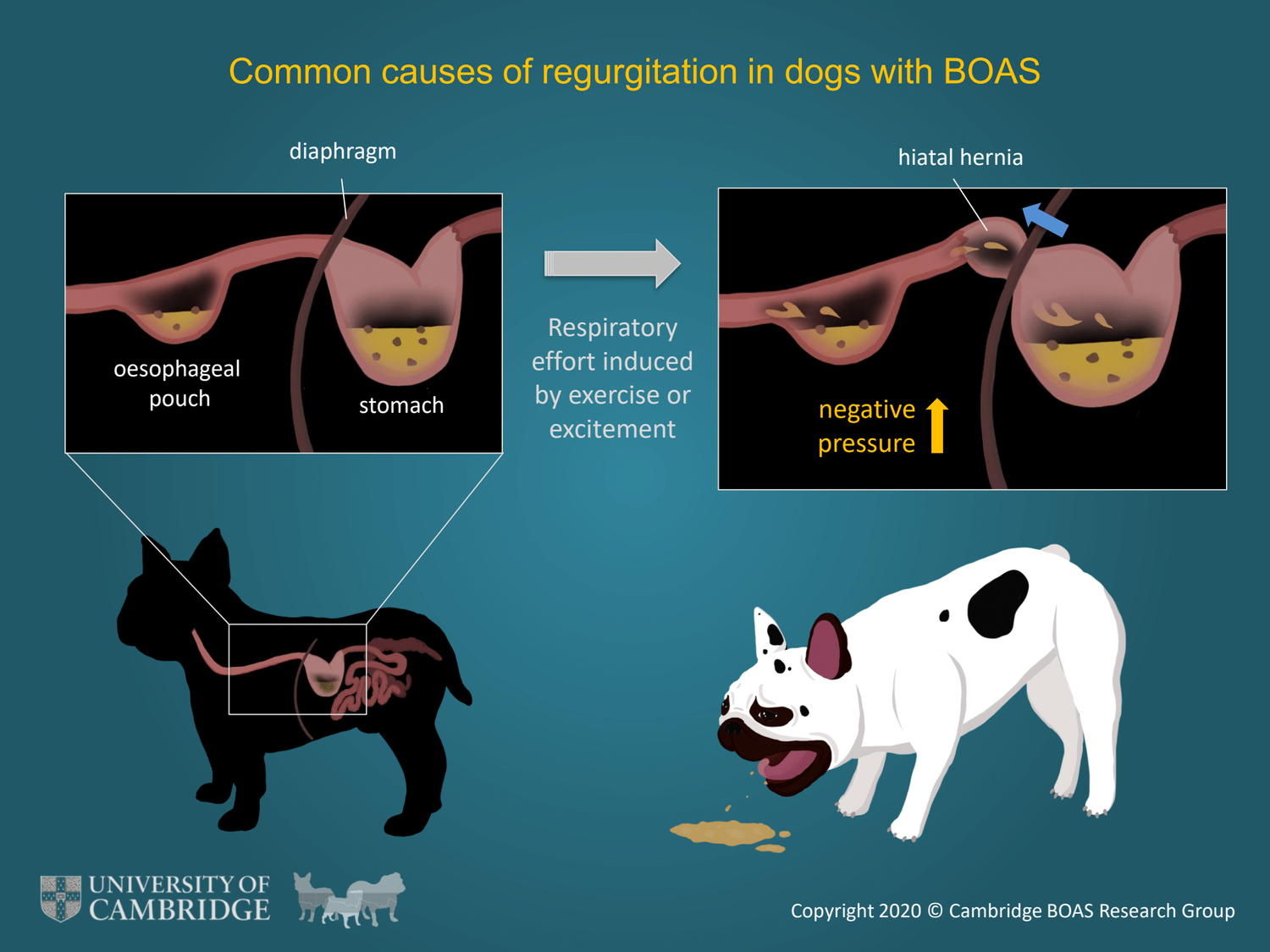
- Monitor for any worsening of breathing signs (e.g. increased breathing noise; sleep apnoea; frequent regurgitation; decreased exercise tolerance) and contact your vet if you are concerned.
Prevention is always better than treatment – however it is not easy to reverse the effects of many generations of selective breeding. We strongly recommend that breeders take responsibility for health testing dogs involved in breeding programmes and that dog owners research the potential diseases within each breed. For dogs with BOAS a combination of surgery and lifestyle modifications are likely to be required to effectively manage their symptoms long-term.
http://www.vet.cam.ac.uk/boas/about-boas/recognition-diagnosis#clinical-assessments
MANAGEMENT OF BRACHYCEPHALIC BREEDS IN HOT WEATHER
Because all brachycephalic breeds have varying degrees of the predisposing anatomical features of airway obstruction, even if it is subclinical, it is appropriate to treat all brachycephalic breeds as having the potential for upper airway obstruction. It is worth remembering that with the shorter face, the less the air will cool before it reaches the lungs.
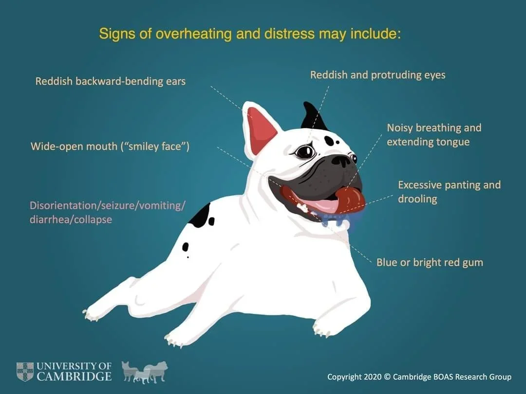
Predisposing Risk Factors - Heat, humidity, exercise, excitement can all increase panting as the dog attempts to loose heat and cool itself – this excessive panting in turn can produce local swelling (oedema) and further airway narrowing, increasing anxiety and body temperature; creating a vicious cycle.
Treatment - If panting hard, cool the dog all over by hosing the dogs down in a bath or a wading pool. Pay particular attention to the head, throat and belly. Do not attempt to make the dog swallow – ice packs placed along the belly, under the throat will help cool the dog – keep going for a minimum of 10-15 minutes, until the respiration rate slows down. If the dog is still having problems, get the dog to the veterinarian as soon as possible. Keep the car air conditioned with the cold air directly in the face of the dog.
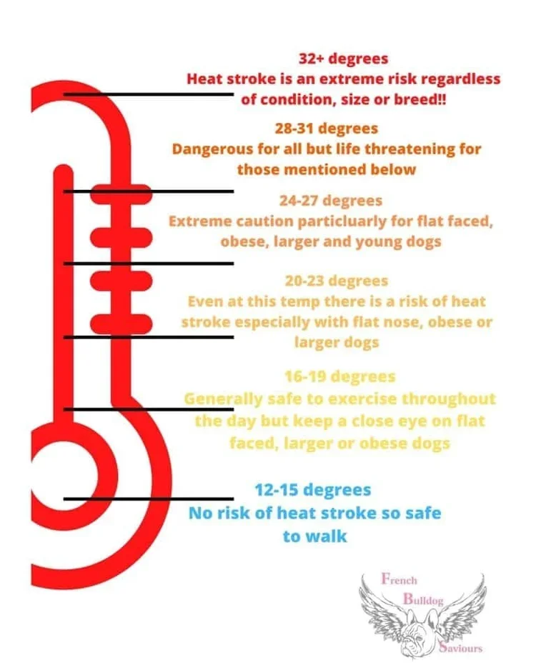
Prevention – be aware of the temperature on a daily basis, weather forecasting generally will give a good idea well ahead of hot weather. Place your dogs on extra electrolytes in their food – this can help them cope with the heat better. Keep your dogs in cool conditions with plenty of through ventilation. Extremely hot weather – the more affected dogs may need to be kept in air conditioning. Fans, wet towels on the floor etc can all be useful items to leave out on hot days.
NARES
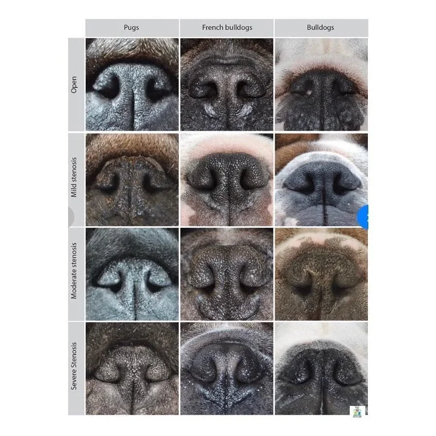
FRENCH BULLDOG NOSTRILS - RECOGNISE WHAT OPEN NOSTRILS SHOULD LOOK LIKE
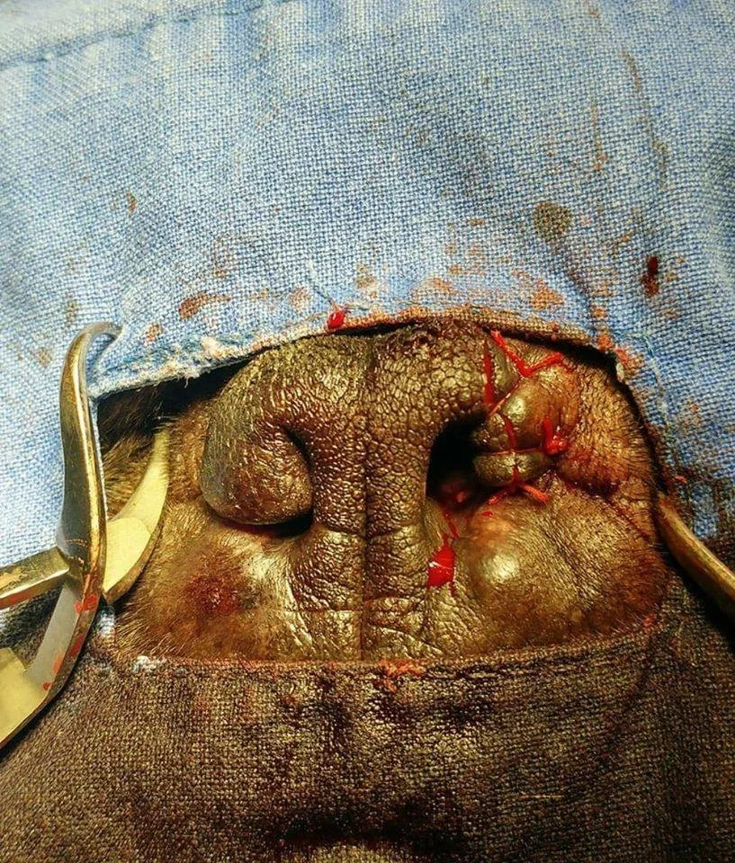
Stenosis has been reported not only in the exterior nostrils which is easily seen, but also in the inner part of the nasal wing (alar folds). As a result, respiratory effort and open-mouth breathing are commonly seen in brachycephalic dogs.
Stenotic nares are considered a risk factor for BOAS, particularly in French bulldogs. French bulldogs with moderate-severe stenosis of nostrils are about 20 times more likely to develop BOAS. Corrective surgery to widen the nostrils is usually recommended for dogs with moderate and severe stenosis. We must strive to breed away from Frenchies with moderate or severe stenosis.
The degrees of nostril stenosis in brachycephalic breeds are defined as follows:
Open nostrils - wide opening.
Mild stenosis - Slight narrowing of the nostrils. When the dog is exercising, the nostril wings move dorso-laterally to open on inspiration.
Moderate stenosis - The dorsal part of the nostril wings touch the nasal septum and the nares are only open at the bottom of the nostrils. When the dog is exercising, the nostril wings are not able to move dorso-laterally and there may be nasal flaring (i.e. muscle contraction around the nose trying to enlarge the nostrils).
Severe stenosis - Nostrils are almost closed. The dog may switch to oral breathing from nasal breathing with very gentle exercise or stress.
HERMIVERTEBRAE
What is Hemivertebrae? Hemivertebrae is a congenital defect in which the vertebrae are deformed, either in such a way that the vertebrae fuse, or that they are wedge-shaped, and cause a twisting of the spine. (show rads of hemivertebrae)
Who gets Hemivertebrae? Hemivertebrae is common in brachiocephalic breeds, such as the English Bulldog, French Bulldog, Pug and Boston Terriers. These breeds are famous for having what is called a “screw tail.” The screw tail results from hemivertebrae in the vertebrae of the tail and is characteristic, even desirable, in these breeds. It can also occur in German Shorthair Pointers and German Shepherds.
What are the Signs of Hemivertebrae? Hemivertebrae of the tail alone is not such a medical concern. However, when it is present in the rest of the spine, it can cause serious clinical signs. Because the deformed vertebrae cause a wedging effect that twists the spine, it can actually cause compression of the spinal cord. Clinical signs include weakness of the hind limbs, pain, urinary incontinence, and fecal incontinence. Signs often progressively get worse, until they plateau at about nine months of age, once the spine stops growing.
How is Hemivertebrae Diagnosed? Hemivertebrae is relatively easy to diagnose using traditional radiographs. More advanced technologies such as myelograms, CT scans, or MRIs are used to detect spinal cord compression.
Why did my Dog get Hemivertebrae? Hemivertebrae is a congenital condition, and those breeds that are selectively bred to have “screw tails” are predisposed to the condition. In the case of German Shorthair Pointers and German Shepherds, it is genetically inherited in an autosomal recessive trait.
How is Hemivertebrae Treated? Many dogs with hemivertebrae show no clinical signs or pain, and do not need to be treated. Rest and anti-inflammatories can be helpful for dogs only mildly affected by the spinal cord compression caused by hemivertebrae. In more severe cases, surgery is necessary to relieve compression on the spinal cord. The procedure used is called a hemilaminectomy, and involves the removal of the intervertebral disc material that is pressing against the spinal cord, followed by stabilization of the spine.
Can Hemivertebrae be Prevented? Because hemivertebrae is a congenital condition, the only way to prevent it is through responsible breeding.
What is the Prognosis for my Dog with Hemivertebrae? Hemivertebrae can vary greatly in severity, from having just one or two abnormal vertebrae and no clinical signs, to having a number of abnormal vertebrae and significant clinical and neurological signs. Prognosis for dogs with hemivertebrae depends on the severity of their case. Decompressive surgery is usually very successful, and most dogs that undergo the procedure regain normal locomotor function.
EYES
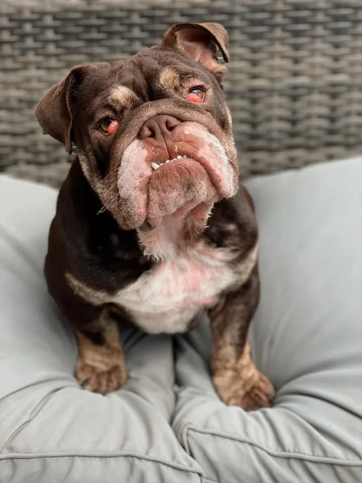
‘Cherry eye’ an everted (rolled out) 3rd eyelid with the gland underneath exposed – this occurs usually secondary to loose eyelids and inflammation of the eye. Usually seen over 6 weeks and under 6 months of age. Low incidence as most Frenchies have tight eyelids.
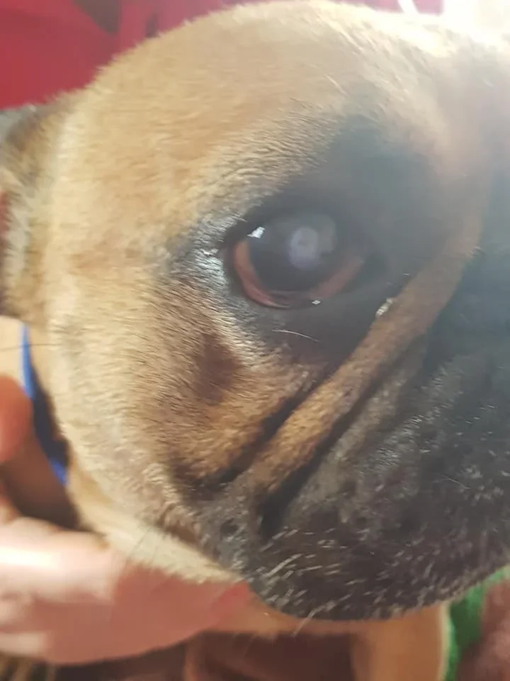
Corneal ulcers are not uncommon as Frenchies age, due partially to the prominent nature of their relatively large eyes. These respond well to treatment, provided it is prompt and effective. Any ulcer that fails to respond to treatment quickly needs to be reassessed frequently by your veterinarian and may require
surgery in the form of a third eyelid flap to rest the eye while it heals.
Pannus – deposition of black pigment on the cornea and subsequent drying (dry eye) of the cornea. Seen in the older Frenchie (8 years and up). This is considered to be an autoimmune condition in many breeds. Once the black pigment starts to deposit on the cornea, usually in the medial edge and accompanied by inflammation on the outer edge of the pigmented area, it cannot be stopped but can be controlled for long periods. Eventually the pigment will cover the entire cornea, resulting in blindness – this process usually takes several years.
Treatment - the condition responds well to the use of cortisone drops and/or Cyclosporin* eye drops.
Liquifilm eye ointment to keep the cornea moist is needed in the more advanced cases. Interestingly, most cases I have seen have also been affected by hypothyroidism. Incidence around 10-15% in the aged Frenchie.


